Biology Lab Techniques
- Title: Introduction to Cellular and Molecular Biology Labs
- Description: Throughout the semester, my lab partner and I were tasked with completing various lab procedures as well as effectively logging all lab information via the use of a lab notebook. The lab notebook and images from my labs can be found below.
- Skills: Throughout my biology labs I have demonstrated a wide variety of lab techniques including microscopy techniques, Gram staining, the creation of standard curves, and SDS-PAGE, among others.
Lab Notebook
Full lab notebook documenting all procedures and results throughout the semester.
Microscopy and Gram Staining
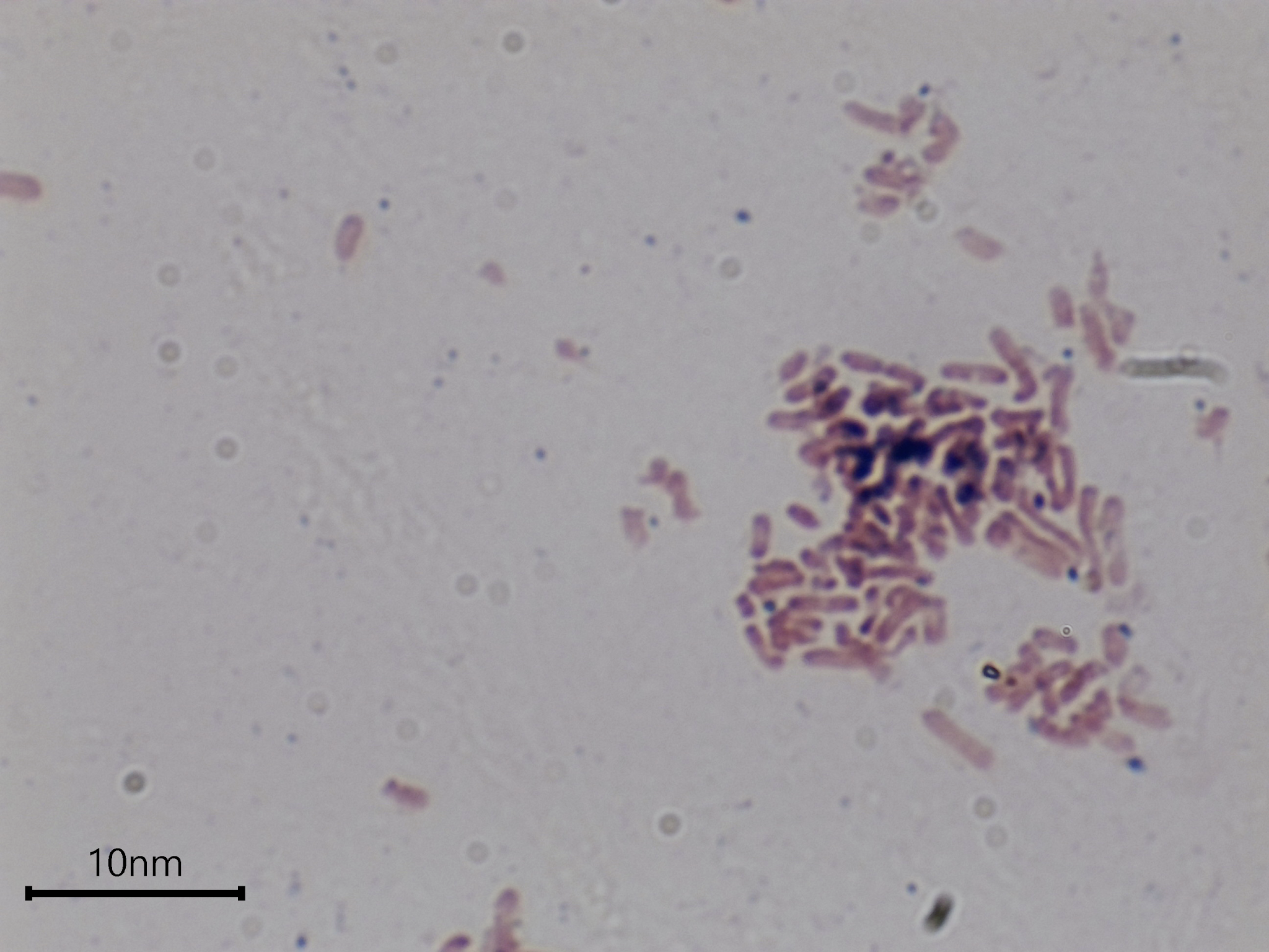
I understand the parts and uses of a bright field microscope, and am able to perform Gram staining techniques in order to visualize the differences in cell wall thickness across specimen. Pictured is the Gram-negative E.coli.
Standard Curve and Micropipetting
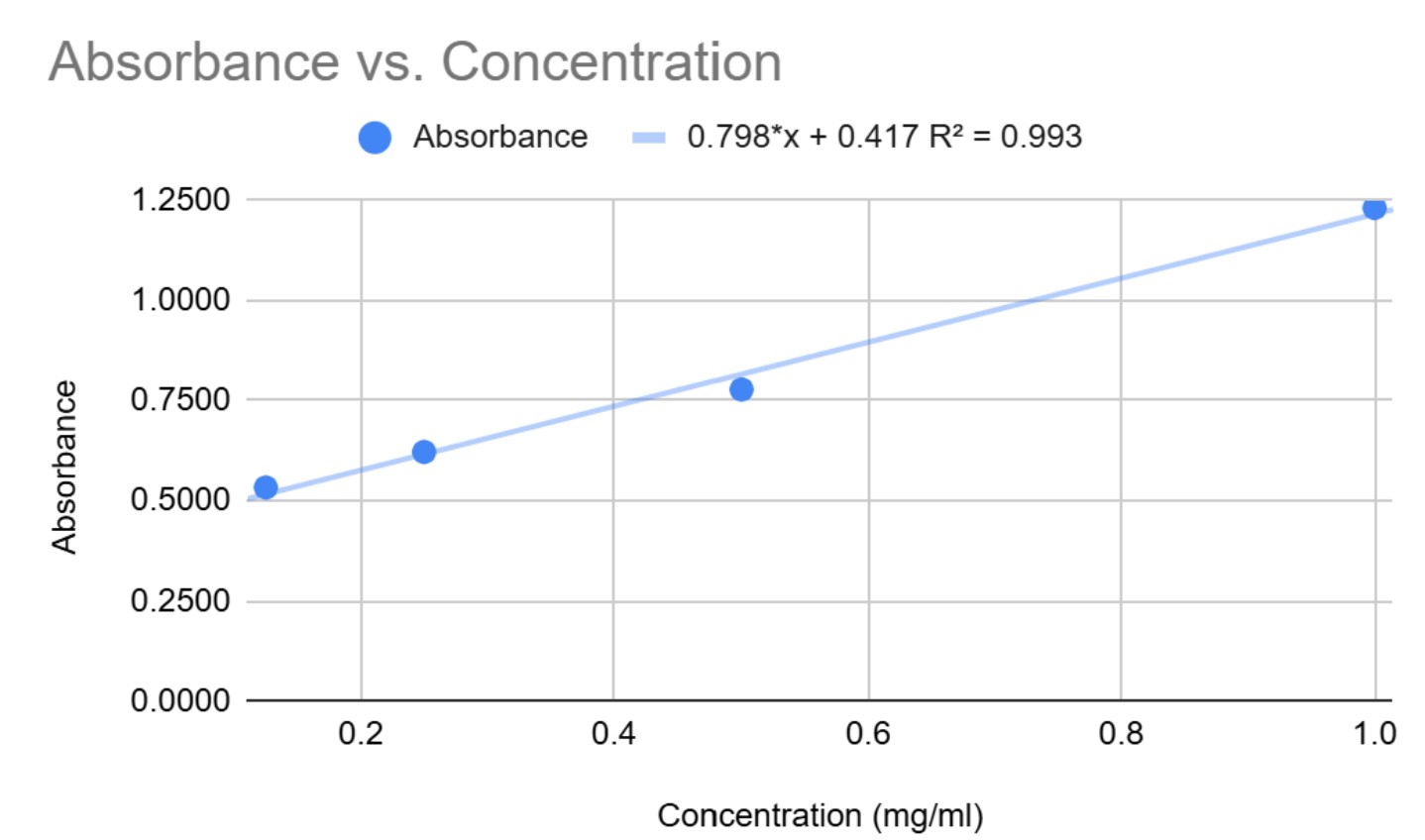
I am able to perform basic micropipetting technique as well as create a standard curve in order to determine an unknown protein concentration. Pictured is the standard curve created for our lab.
Enzyme Kinetics
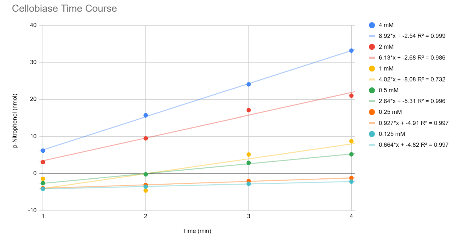
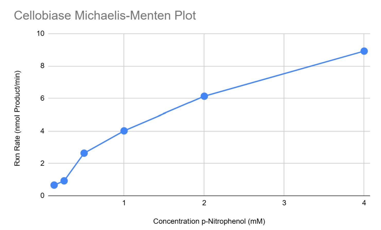
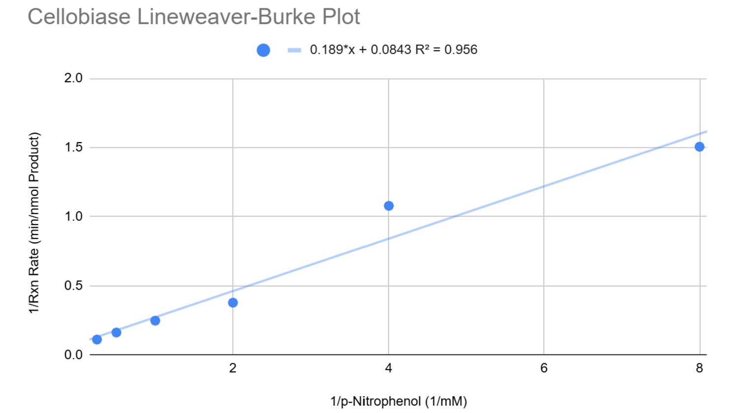
I understand enzyme kinetics and am able to perform time course reactions. Additionally, I am able to compile data in order to create time course, Michaelis-Menten, and Lineweaver-Burke graphs. If I were to redo this lab, I would redo the reactions for the 1 mM concentration since the R² value was less than ideal.
Western Blot and SDS-PAGE
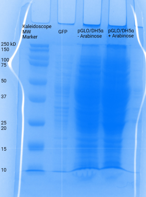
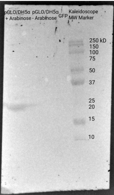
I am able to perform a Western Blot and SDS-PAGE. I did not see a band in the GFP lane like I should have. If I were to do this experiment again, I would make sure that the GFP was put into the gel correctly since the GFP lanes were incorrect in both the SDS-PAGE and Western Blot.
PCR Motility Plate
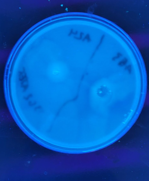
I am able to create a PCR motility plate. Pictured at the left is the A24 motile E.coli. At the right is the A53 non-motile E.coli.
Phase-Contrast Microscopy
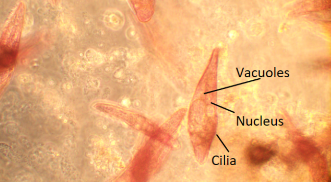
I am able to utilize a phase-contrast microscope in order to view living specimen. Pictured is Blepharisma at 200X magnification.
Fluorescence Microscopy
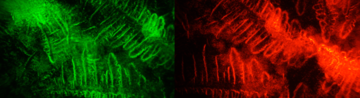
I am able to utilize fluorescence microscopy in order to view a variety of different specimen. Pictured is a GFP and m.Cherry E.coli mixture under green and red fluorescence.